Chapter 1:
The Quick Guide to Understanding How Your Knee Works

The knee isn’t super complex. It bends, it straightens, and allows for a bit of rotation. Yet even simple things sometimes need a user manual.

That’s because a pair of healthy knees is essential to active living. Try getting off the floor or landing with your legs locked out to understand why.

Without the knees walking and running would be awkward and much slower, jumping wouldn’t be much of anything, and our ability to raise and lower ourselves to a chair or ground level would be extremely limited. They also dissipate a ton of force which lessens the loads on our hips and low back. 

So thankfully we have them, even if they are prone to pain and injury. 

This is partly due to restrictions on how the knee is designed to move. Apply enough force the wrong way and you will quickly be out of commission. 

It also plays the middle man between the ankle and the hip. Often taking the brunt of any movement issues coming from these areas above and below the joint even with everything around the knee working just fine.

We’re here to help guide you through these issues, but the first step is to develop an understanding of your knees. From there it should make more sense as to why they hurt and the plan to fix them.  

This article is the 101 to the knee joint. We’ll walk through the major structures around the knee, laying the groundwork to get deeper into the issues that affect it and ways to fix it.

Bones of the Knee- “The Framework”

Let’s start by reviewing the bones that make up the knee. There are only 3 (and kind of a 4th) that lay the groundwork.

Femur

(aka- Thigh Bone)

The femur is the only bone of the upper leg. It is both the longest and strongest bone in your body and is excellent as at absorbing force.

It’s not pictured above, but as you’re probably aware, the upper end of the femur connects to your hip as a ball-and-socket joint. Thus, movement at the hip joint directly affects the position and alignment of the knee.


Tibia/ Fibula

(aka- Shin Bone)

The tibia is your shinbone and creates the lower part of your knee. On top, the tibia is a shallow surface where the cushioning for the knee sits. This cushion is called the meniscus and will get further discussion later.

Some people say the fibula is a part of the knee joint too; however, most don’t agree since it doesn’t directly connect to the knee. It’s still worth noting since it lives so close and is the site for an important ligament attachment for the knee.


Patella

(aka- Knee Cap)

The patella is best known as the kneecap and sits over the top of the knee in a groove in the femur simply called the patellofemoral groove.  It acts as a hard hat to protect the inner workings of the knee joint, but most importantly it acts as pulley system to help straighten the leg.


The Patella Pulley System

To further explain the pulley system of the knee joint, think of the muscles on the front side of the leg as a rope, which pulls over the patella. The patella provides mechanical advantage to help increase the amount of power that the quadriceps can generate.

You can see from the picture that without the kneecap, the quad tendon would essentially pulling on the lower leg from directly above it. This would make it really tough to extend the leg, especially from positions where the joint was extremely flexed, like crouching.

This also highlights the issues that come up if the knee cap isn’t moving correctly. If the patella isn’t tracking the right way in its grooves, it will affect how knee moves and limit the legs ability to generate force.

Ligaments, Tendons, & Cartilage- “Connecting the Bones”

The next piece of the puzzle is what brings these bones together. Just like our other joints, it’s a connection made of ligaments, tendons, and cartilage.

Many of these structures have infamous names for their association with major knee issues. Things like the ACL, meniscus, and patellar tendon probably sound familiar, but ever wonder what they are?


Ligaments- Connecting Bone to Bone

Starting with the ligaments, which are the the things that connect the bones together.  Remember, ligaments connect bone to bone. Each of the ligaments has 1-2 specific jobs, for example, stopping the lower leg from sliding too far forward.

There are four main ligaments in the knee:

ACL- anterior cruciate ligament
PCL- posterior cruciate ligament
LCL – lateral collateral ligament
MCL- medial collateral ligament

The ACL and PCL are termed the “cruciate” ligaments for the cross, or x-shape, that they form inside the knee joint.

  • Patellar and Quad Tendon
    The quad tendon is located on the top of the knee, attaches the quad to the patella.    Go below the knee cap and you’ll find the patellar tendon which isn’t actually named correctly.  It’s actually a ligament (remember that means bone to bone) because it attaches the kneecap to the shin bone. It’s common for both of these areas to become irritated, resulting in forms of tendonitis, usually due to strain and overuse.
  • Hamstring Tendon
    The hamstring tendon crosses the back of the knee and attaches to the tibia and fibula. Some will have irritation here for various reasons, but it’s more common to strain a hamstring near the hip rather than the knee. You’ve probably heard of an athlete having a high-hamsting strain. For another fun knee fact, one of the hamstring tendons is a popular replacement for then ACL  during surgical reconstructions.

Cartilage- “The Cushion”

  • Meniscus
    The largest piece of cartilage in the knee is called the meniscus. The meniscus is a C shaped ring that lives on top of the tibia (shin bone). It helps cushion a lot of force coming from the ground and reduces friction.You can see why the meniscus is easy to injure. It is the cartilage that helps take the force that is coming from the ground and disperse it.The meniscus has been likened to a sponge in that it needs some water to be healthy (we keep water in the meniscus with the right level of impact). But, too much impact (or too much water in the sponge) can leave the sponge brittle.
  • The Other Cartilage
    There is lesser-known cartilage to the knee that’s equally as important called articular cartilage.Articular cartilage covers the bone ends to reduce friction when bones rub against each other. Most importantly it covers the areas between the femur and tibia, and underneath the kneecap.Different than other areas of cartilage, this cartilage is not great at absorbing force, and instead more helpful for reducing friction.

Muscles and Bands – “The Movers”

Now we have all reviewed the makeup of the knee, let’s talk about how it moves.

Things that Move the Knee

These are the major movers that live around the knee joint. This means that when they contract they have the direct ability to make the knee move. This isn’t a comprehensive list, but covers the major players:

  • Quadriceps (AKA quads)
    The quads are made up of four different muscles, they are really good at getting our knees straight, and doing it with a lot of power
  • Hamstrings
    The hamstrings are made up of three different muscles. They bend our knees towards our butts. They are also a big dynamic stabilizer of the knee. Meaning they help support the knee when things are moving quickly.
  • Illiotibial band (AKA IT band)
    The IT band starts at the hip and attaches outside the lower part of the knee.  Although as the name implies, the IT Band is not a muscle, it’s is a band of fascia, but it’s appropriate to put it here because it’s an extension of a muscle and it’s often treated like one. We’re inclined to smash, roll, scrape, and stretch it to “loosen” it up, but you will not change the very strong fascia of the IT Band.
  • Adductors (AKA groin muscles)
    There are also three groin muscles that pull out leg towards the midline.
  • Gastrocnemius (AKA calf)
    You may have never thought of the calf behind a part of the knee, but the way our calf muscles attach to the leg it will also help bend the knee.

Stability

Taking a step back and looking at the big picture we can see how important the knee is, but linking the pieces together we can’t forget about what happens above and below the joint.

The stability that comes from the ankle and the hip directly impact how the knee works.

It should make sense though. Now that we have reviewed the bones and muscles that make up the knee, you may have noticed some overlap between the bones of the knee and bones of the hip or ankle. That is because they are the same.

For example, the top part of the knee joint (the femur) is the same bone in our hips. So it can be easy to see how what happens at the hip directly affects the knees.

We won’t dig into the specifics of how these are intertwined in this article, but understanding how closely these joints are related can be helpful to improving the stability of the knee by looking above and below the joint.

From here we’ll start laying out specific pain points that affect the knee, but more importantly, how to fix them.

Chapter 2:
Guide to Meniscus Tear Recovery

This article will be your guide to meniscus tear recovery and hopefully provide some clarity on this common knee issue.  We’ll start with an easy understanding of the meniscus and what it does. Then we’ll help you identify potential issues and what research says about the most current treatment options.

The first note is that not all meniscus tears are the same and management will likely look a bit different for each case.  Nonetheless, with a better understanding of your meniscus, you can help navigate the best treatment option for you.

Quick Facts About the Meniscus

The Meniscus Picture

As we reviewed in The Quick Guide to Understanding How Your Knee Works, the knee bends and straightens like the hinge on a door.  Inside the hinge are two “C” shaped cartilage rings called the meniscus.  


It’s easily termed medial meniscus for the inner ring, and lateral meniscus for the outer ring.  Together they create a bowl for the femur to sit.  It helps provide stability for the knee and disperses loads evenly throughout the joint. It also reduces friction and adds padding to protect the bones from rubbing against each other.


When the doc says “bone-on-bone,” they’re trying to explain that the meniscus padding (along with other cartilage) is missing.  But the impression that the body is worn out like old brake pads on a car isn’t accurate and may do more harm than good.

It implies there is no way to fix the issue without a repair and the problem will only get worse.  Yet for many issues, including meniscus tears, a plan of exercise and rest often works well and is an ideal first approach (ref).

How Your Meniscus Becomes Damaged

Meniscus tears can occur in nearly any individual— ranging from elite athletes to older adults. Despite the different demands, abuse to the meniscus usually comes from landing and changing directions.

To help explain this, think of the meniscus as a wet paper towel squished between two rocks. If you merely compress the towel between the rocks nothing will happen. However, if you squish and twist the rocks, you can imagine how it would like the tear the paper towel. 

For older adults, tears develop with much less force. Think of the same wet paper towel analogy, but using a thin towel rather than a thick super-absorbent one. The thinner towel won’t take as much force to tear. Depending on the quality of the meniscus, walking stairs, or sitting or standing from a chair, could be enough force to tear the meniscus.

All of this makes the meniscus seem all too fragile— but it’s far more robust than a wet paper towel. It’s thick and rubbery, built to take a beating. Thus, not everyone with knee pain has a meniscus tear, nor does a tear even mean you’ll have pain. 

When Things Go Wrong 

Depending on the type, degree, and location, people experience different symptoms.  Below you will see the most common types of meniscus tears.  The upper row is a milder version than the more progressed version under it.


Additionally, a traumatic tear in a young soccer player will feel and recover much different than general wear and tear that’s common in people over the age of 40.


Yet, the common symptoms associated with a meniscus tear include:

  • Pain with Clicking*
  • Swelling that progressively increases over 24 hours 
  • There may be limited ability to straighten the leg completely or bend it all back all the way depending on the location of the tear.

(*Important Note- Many people have clicking in their knees that means nothing, so don’t freak out over the little creaks and pops you get in your joint.)

The gold standard for diagnosing a meniscus tear is by MRI. There are also clinical tests that can hint at a tear, which you can check out one in the video below. You can see it’s an aggressive test that could further your issue, so this is best left to a trained professional.

Acute vs. Chronic Meniscus Tears

The initial question a medical professional will often have about your injury is, “How did it happen?”  

For many tears, it occurs from a sudden or specific event such as a hard cut or land, and many report hearing a pop.  Despite the nature of the injury, most are still able to keep moving and many athletes will continue to compete in their event.  It’s not until later when they notice swelling, pain, or tightness, do they realize there is an issue. 

Meniscus tears due to accident or injury may also involve damage to other stabilizers as well.

“The unhappy triad” is the unfortunate, yet common situation, where the meniscus, anterior cruciate ligament (ACL), and medial collateral ligament (MCL) are torn during a fall, hit, or twist.  It usually occurs with force moving from the outside of the leg inwards with the foot fixed on the ground. All three structures are critical components to the stability of the knee, thus it’s a different issue altogether than just a damaged meniscus.

The other type of meniscus tear is more chronic in nature, without a cause or explanation.  These tears are more likely in people who have arthritis in the joint already. We’ll get more into treatment later, but many times chronic tears don’t make good surgical candidates due to complications from underlying arthritis in the joint (ref).  

Blood Flow

Meniscus tears also differ based on their location within the ring.  The “red-red” zone is the outermost part of the ring and has the best blood flow.  This allows for increased nutrients and metabolites to give the tissue the best opportunity to heal.  The middle section is the “red-white” zone and does not have direct blood flow, but is close enough to the red-red zone to get some trickle over. The white-white zone is the innermost portion of the meniscus ring and has the least amount of blood flow.  Tears in this area can heal, but it’s much less likely.


Treating Your Meniscus Tear

The good news is many meniscus tears will heal on their own.  Based on what we currently know, the location of the damage in either the red-red zone vs. white-white zone is most telling for the tear’s ability to heal.

Yet some factors make surgery a necessary option.  Sometimes a meniscus tear can act like a hangnail, constantly getting caught up and aggravating things.  Other times, the tear may not be severe, but in a location that gets extra abuse. Like the neverending cut on the finger that’s constantly getting bent, bumped, and reopened 

The Conservative Approach

The difficulty is that you won’t know your opportunity for rehab success without giving it a try.  A physician can make an educated guess, based on imaging and your history, but this is no guarantee.

Acute and or painful tears usually start with a period of rest to allow things to calm down,  followed by physical therapy for 4-6 weeks to improve strength and range of motion.

Some patients will find that their tear won’t afford them the option of conservative treatment.  For example, for a big flap that’s causing locking and severely limiting the range of motion point to surgery as the only option.

Other Potential Aids (That Aren’t Surgery)

Cortisone is a steroid injection into the location of the tear. Cortisone will NOT heal the meniscus, but reduces pain for 3-6 months for more effective stretching and strengthening. 

It’s helpful for taking the sting out of the issue, but won’t likely have any lasting effect without strengthening.  And the most significant trouble with cortisone is the lack of short and long term research on its use.  

PRP (Platelet-rich plasma) is a newer treatment option and a hot-button topic.  The short story is a mixture of your own blood cells is injected into the injury location to promote the healing process. 

Studies on PRP for the meniscus have been promising but not definitive (ref).  The red-red zone appears to be an effective target for PRP, while treatment in the white-white zone seems to be less effective. Again, the red-red zone heals the best because it has the best blood flow to the area. 

Ultimately PRP needs more research. We currently don’t have a great answer for who it works for, making it a system of guess and check.  Insurance is not reimbursing for it yet either, but if you’ve got a couple of hundred dollars lying around, it may be worth it.

Surgical Options

If the conservative options don’t work, there are two surgery types to help correct the issue.

Surgical Removal

One option, called debridement, involves the surgical removal of the torn area of the meniscus.  This is a minimally invasive procedure using a small camera and tools inserted into the joint via keyhole incisions. Typically individuals return to their desired activities between 6-8 weeks. 

Surgical Repair 

Or it’s possible to repair the meniscus rather than cutting out.  This is an arthroscopic procedure as well, using a small stitch to connect the meniscus back to itself.  

Although, this is a delicate process that needs careful management afterward.  Some surgeons describe the procedure as sewing tissue paper back together.  

Depending on what research you are reading, the recovery for this process can take anywhere from 3 months to full year, but the big take-home is that it’s much lengthier than simply cutting it out.

Moving Forward with Your Meniscus Tear Recovery

At this point, this is what many of you are thinking…

I can give rehab a try, but won’t know if it will work for several weeks.

Or, I could have it stitched back together, and be out up to one year.

Or, I could have a doctor cut it out, and go right back to my activities in just a couple of months. 

While the third option probably sounds the most appealing, looking past the 6-8 week recovery, there are long term consequences to consider for removing a piece of the meniscus.

Going back to the original function of the meniscus, its job to help disperse loads within the knee. Removing rather than repairing a torn meniscus leaves more contact of bone to bone, which increases the chance of developing arthritis later in life.

Ultimately it’s worth the time investment if the meniscus can be saved (ref). 

Initially, try conservative management—and I mean seriously try—where you give your knee the best chance to rest and recover properly. Even if it’s not the ultimate fix for your knee, you only lost a few weeks and set yourself up to come back better and stronger after your procedure.

If you decide on the surgical route, it’s worth discussing with your surgeon about debridement vs. repair, and how it will change your rehabilitation process and the long term health of your knees.

Conclusion

Getting past a torn meniscus is not a one-size-fits-all solution. There are many options for managing a meniscus tear.  Hopefully, this laid out some of the information to help you make a more informed decision going forward!

Chapter 3:
A Quick Guide to Knee Sprain Rehab

A quick turn followed by a pop of the knee may be the pinnacle of oh s*** moments. It’s a common occurrence in sporting events but also happens in everyday life.

It’s not always a grave situation—knees make creaky noises all the time and are quite robust— but the dreaded knee sprain does happen to many and is immediately impactful.

Despite the suddenness of the injury, it leaves a prolonged aftermath. Not only painful and limiting, but the biggest struggle is the extended time to heal, and potentially the need for surgery.

In this article, we’ll help explain the knee sprain and guide a path to getting you back into action.

Anatomy Review

A knee sprain is the result of damage to the ligaments of the knee joint. There are several major ligaments, and a few lesser ones, that will be covered shortly.

A ligament is a fibrous tissue that attaches bone to bone. Ligaments are often confused with tendons, but it’s a different part of the anatomy.

If you want a full review of the knee and how everything ties together, check out our Quick Guide to Understanding How the Knee Works. But if you want to get straight to it, let’s focus specifically on the ligaments of the knee.


Understanding the difference between the types of tissue in the knee is important because it helps answer the usual question…

 “How long will this take to heal?!!?”

The reason is due to blood flow! In general, the ligaments in our bodies have a poor blood supply. Bone has a great blood supply, the muscle has a great blood supply, but cartilage, tendons, and ligaments do not.

Without quick access to new blood, and therefore healing factors that our body uses to repair, it takes an extended time to heal those strained or sprained tissues.

Sprain vs. Strain vs. Tear

Damage to the ligaments gets classified as either:

A sprain is when we overextend a ligament.

A tear is when we overextend, and the two pieces lose their connection.

(Note- A strain is another common orthopedic injury term, but this occurs when we overextend a tendon or muscle.)

All ligaments have a threshold of strength. Just like a rope has an amount of force that it can withstand when holding two objects together. And just like the rope, if a ligament gets stretched too quickly OR too hard, then it gets damaged.

Each ligament provides a specific restraint to prevent the knee from going too far in one direction. Here are the major ones and usual suspects when it comes to knee sprains:

  • Anterior cruciate ligament (ACL) is commonly injured in fields sports, volleyball, gymnastics, and skiing. Typically injuries occur by a hit to the side of the knee, or if there is a twisting motion with hyperextension at the knee.
  • Posterior cruciate ligament (PCL) is most commonly injured in what is called the “dashboard” mechanism—when knees are bent to the chest (called hyperflexion) and the shin bone is pushed backward.  This happens in a car crash (hence the name) or may also occur by getting tackled while the knee is bent.
  • Medial collateral ligament (MCL) commonly injured in field and ice sports. This ligament protects the knee from bending inwards.
  • Lateral collateral ligament (LCL) protects the knee from going out and is injured much less than the MCL.
  • Medial patellofemoral ligament (MPFL) is a small but mighty ligament that helps to stabilize the knee cap in the groove over the knee. An injury to the MPFL is likely anytime there is a dislocation of the knee cap.

You have other ligaments in the knee, but you’re not as likely to injure them.

Grading Your Sprain

Sprains are graded by the level of injury to the tissue. These grading systems vary for different ligaments in the body, but the most common scale for the knee ligament injuries is I-III (least to worst.)

A grade I sprain occurs due to overstretching. It is considered a “mild” sprain and usually results in minor swelling and stiffness of the knee. Current evidence shows that a grade I sprain in the knee will heal and return to normal in anywhere from 4-8 weeks.

It’s a frustratingly long time for some “minor” stretching. But the blood supply issue pushes the healing time out longer than most people suspect. And since the knee is so crucial to getting around, even minor discomfort doesn’t go unnoticed.

A Grade II sprain means there is a small tear, but it’s not all the way through the ligament. At this time, it’s unknown how much those sprains truly heal, but it’s safe to assume that you can get back to your sport after some time off. 

A Grade III sprain is equivalent to a full tear in which the ligament has torn apart from itself and don’t expect these to heal. Grade III sprains often undergo surgery, although there are currently many valid non-operative options.

Diagnosing Your Knee Sprain

If you’ve been hit with a knee sprain, start by looking for the following Red Flags as a reason to seek further medical evaluation.

Medical Red Flags

  • Knee gets “locked” in position- either bent or straight
  • New onset of painful clicking or catching
  • Feeling of instability or giving out
  • Swelling that lasts >5 days

If you test out of all these and want to save the time and money, then it’s fine to wait it out.  

But, if you’re worried about your knee, get it checked out by a medical professional for peace of mind.  It’s not really an emergency situation though, so no need to rush to the emergency room.  

If you have direct access to a physical therapist in your state, this is probably your best resource for the sake of time, cost, and a more comprehensive recovery plan when appropriate. Otherwise, a general practitioner or urgent care clinic can evaluate the issue and provide further recommendations, which are usually rest and anti-inflammatories, and potentially a referral for a sports medicine doctor (ref).

The Process for Knee Sprain Rehab

If you’ve sprained your knee, the first question is likely: What should be done? It’s a simple, yet challenging task, and that is resting the injury.

Rest will be slightly different for each type of sprain, but in general, it means eliminating painful activities. In the best-case scenario, inflammation takes about 14 days to resolve. Thus, the rule of “if it hurts, don’t do it” is a safe bet to follow for 2 weeks. Trying to push through the pain will only bring further inflammation and ultimately slow healing time.

In total, you’re looking at a healing time of 6-8 weeks, but will be feeling better between 2-4 weeks. You will notice a reduction in swelling, and your range of motion will return to normal. You’ll feel that it’s time to get back to life—unfortunately, it’s not done healing.

It’s in the window of weeks 2-4 that people run into issues because they jump right back into their previous activity level. Even though the knee feels better, it’s still missing some stability, which increases the risk of hurting the same ligament again. If not something worse!

It’s during these weeks that a properly structured exercise plan becomes super important.

Low impact exercises targeting the glutes, core, and leg muscles will keep those muscles engaged, while healing occurs. This makes it easier to return to full activity, and potentially even solving some of the underlying strength issues that caused the knee sprain to happen.

If you need help with this, we walk you through everything in our 30-Day Knee Fix.


Preventing Knee Sprains

As Ben Franklin once said, “An ounce of prevention is worth a pound of cure.” And considering the time and limitation caused by a knee sprain, an effort towards prevention is worth it. That’s especially true if you’re at higher risk, like in a sport with lots of contact or cutting.

When it comes to knee sprains, there are some things you can’t control. These are known as non-modifiable risk factors and include items such as:

  • Ligament Size (some people just have smaller ligaments)
  • Gender (women are at higher risk of knee sprains)
  • Physical requirements of a sport or task (for example, soccer players are at higher risk of knee sprains than bowlers.)
  • Background or history of training (previous time spent working with a strength coach)

Yet there are still things you can do to reduce the risk of knee sprains.

Strength

Building the strength of your muscles can help reduce the risk of a knee injury.

The knee is a rather simple joint—which flexes and extends—using the ligaments to keep it along its tracks. A blow to the knee can knock it out of place damaging the ligaments, or forces of just bodyweight moving in an awkward direction can strain a ligament as well.

That’s where strength and stability come into play for protecting the knee joint. Strong muscles slow the body and effectively transfer forces.

For example, the picture above shows the common mechanics for non-contact ACL tears. Someone with weak external rotators is 8x more likely to injure their ACL than someone who isn’t (ref). That’s because the external rotators (AKA- the glutes) help to control the position of the knee when landing and changing direction without overloading the ACL.

Speed and Timing

Again, speed is an essential component behind injuries to the ligaments as well. You could demonstrate the above position slowly and have no risk of injury, but if you got pushed that way, things could easily go wrong.

Again, we prevent entering these positions through the control of the hip extensors, abductors, and extensors. But it’s not enough to just have the strength of the muscles, it’s also essential to engage them quickly and with the right timing.

For that reason, practice and training of explosive cutting and jumping in a controlled situation helps prepare athletes to safely take on the demands of their sport. In sports performance training, this is commonly known as plyometrics, but it’s important for more than just sports performance.

For Now and Forever

If you landed on this to help get past your knee sprain, I hope you found the answers you were looking for. It’s going to take some time, and we would love to help guide you through that.

Our 30-Day Knee Fix will give you a progression of strengthening and active rest to get you back to where you once were.

But it doesn’t end there!  Hip and Core strengthening is an important part of knee injury prevention and should be part of every athlete and active person’s regiment.   Be sure to check out our glute and core guide to learn more about the key muscles needed to fight off injury.

Chapter 4:
The Crossover Approach to IT Band Syndrome

Here we’ll outline an IT band syndrome treatment plan to help get you moving pain free again.

IT band syndrome plagues the outside portion of the knee and is one of the most common forms of knee pain. The problem usually arises as an overuse injury, especially after ramping up the number of knee bends in a day. The obvious culprits are running and biking, but those aren’t the only activities that suddenly spike in volume. Jumping into a workout plan with more squats and lunges than you’ve done in a lifetime, or rolling out of bed and deciding it’s a good day for some serious hiking, are other examples of ways you might make your knee cranky.

The symptoms are usually subtle or non-existent to begin. Many runners say they’re able to start their runs pain-free, but shortly into it, a sharp pain develops to the side of their knee — the feeling of being stabbed by a knife.

Many assume it’s an issue coming from tightness, prompting stretching and foam rolling of the outer thigh. This idea was once strongly supported by articles and videos on “breaking up trigger points” or “releasing” the IT band from professionals on the internet. As of late, there has been an outcry against this form of treatment, since it doesn’t do much (although we’ll show you later that it still may be helpful.)

What the evidence has found is that the IT band is much too dense to make it softer or longer. Rubbing on your thigh isn’t harmful and probably feels good while you’re at it, but unfortunately, it’s not tackling any underlying issues and is, therefore, not much of a long term fix.

In this article, we’ll take a good look at the science on IT Band Syndrome and hopefully give you some confidence to get around it.

Anatomy & Function of the IT Band

A general anatomy refresher on the knee will make this article a bit clearer. You can do that here in The Quick Guide to Understanding How Your Knee Works.

Taking it a step further, examine the IT band, or more precisely called the iliotibial band.

(Flato R, Passanante GJ, Skalski MR, Patel DB, White EA, Matcuk GR Jr. The iliotibial tract: imaging, anatomy, injuries, and other pathology. Skeletal Radiol. 2017;46(5):605–622.)

It’s a thick band of fascia, that intertwines with other fascia starting along the crest of the hip. It then runs along the outside of the leg where it finally attaches to the outer portion of the knee. Along the way, it’s a connecting place for the tensor fasciae latae, or TFL for short, on the front side, and the gluteus maximus muscle on the backside.

It covers a lot of distance, spanning both the hip and knee joint, leading many to assume any pain along the outside of the hip or thigh is some form of IT Band Syndrome. But those are different problems.

IT Band Syndrome a condition tied to a pinpoint spot outside the knee where the IT Band runs over a bony place called the lateral femoral condyle. The long-held belief is that irritation was coming from the IT band rubbing over this bony notch, but now we aren’t so certain (ref).

A 2006 study introduced evidence that the IT band is anchored down too well to roll around and get hung-up on things. Instead, they believed the irritation was more likely from a fat pad, a bursa, or other connective tissue getting compressed under the IT band (ref).

A later study partly refuted this. Saying the IT band does roll along the outside of the knee but didn’t conclude either way if it was an issue of friction or compression (ref). Surprisingly this is where we stand on the IT Band after more than a decade of research.

Regardless of the underlying problem, unless you’re looking to cut something out, you’ll approach it the same way.

Diagnosis

Before we get too carried away, it’s a good idea to determine if IT Band Syndrome is what you’re dealing with.

It’s usually a straightforward diagnosis rarely requiring imaging or even special tests. Pain in the right spot, paired with a recent increase in activity, and no injuries to the knee, point to IT band syndrome. But there are other issues, like the strains to the LCL or a meniscus tear, that may present as an IT band issue.  Our Red Flag Screen is a good evaluation of these other major issues.  Reach out at support@crossoversymmetry.com and we’ll walk you through it.

Unfortunately, the treatment may be lengthy, but with active participation— things like activity modification, stretching and strengthening— most are able to navigate the issue without any advanced treatment.

IT Band Syndrome Treatment

Here’s your game plan to help treat IT band Syndrome.

Rest

I’m sorry to say…this will require rest. It’s the four-letter word that every endurance or workout junkie hates to hear. They also happen to be the ones who will get IT band syndrome.

The unfortunate truth is that stopping provocative activities, like running and cycling, is the most important criterion to calm it down. An interesting side note related to rest and chronic use injuries like IT band syndrome is that many times surgical procedures work simply because it forces the issue of rest.

Start with a timeline of 2-weeks off from running, biking, or anything else that involves repeated flexion and extension of the knee. If you’re looking to get your cardio fix, give swimming a try. Or some runners find that they can bike without provoking symptoms.

After 2-weeks off evaluate the progress and your ability to move forward.

The downtime is relatively easy to navigate and fill with other activities. The real problem and where many hit setbacks is the return to activity.

The usual story is a dedicated runner starts struggling with knee pain. They take time off and do all the rehab stuff, then excitedly wake up one morning feeling better than ever. The happy ending is then ruined when they pick up right where they left off and piss off their knee again.

It’s important to return to activity through graded exposure. After 2-weeks off, take another 2-weeks as an on-ramp to getting back to where you once were. This helps reacclimate the tissues to stress and helps you gauge what you’re truly ready for. Just hit the eject button if you feel things get worse.

Pain Management

Most people with IT band syndrome have mild discomfort throughout the day. Simply backing off activity is probably the best way to deal with the pain issue.

Still, some might find it restricts them from activities and disrupts their rest. In this case, NSAIDs may be useful to fight off pain. But when returning to activity, logic would say it’s best to see how the body responds without any pain inhibitors.

Then for the last resort, corticosteroid injections may be considered, and that’s really only if visible swelling is present.

The Rehab Plan

Unfortunately, there is no definitive cure to IT band syndrome other than time to allow things to calm down. Still, additional work may help speed the process and keep pain from returning later on.

There are two main theories behind the treatment strategies. Tackle the IT band itself or the things that work closely with the IT band.

For the former, your IT band is not going to change by your own doing. The IT band is a thick fibrous strap of tissue, akin to a steel cable running down your thigh. Any effort to stretch, mash, or strengthen won’t change your IT band (ref).

What you can change are the muscles that tension the IT band and your biomechanics. The following are the common exercise recommendations to help fix your knee issue.

Runner’s Program for IT Band Syndrome

Our 30 Day Knee Fix helps guide this graded exposure with activity restrictions that progress over the one-month program. Here are our recommendations for runners dealing with knee issues:

Along with this, it’s advisable to avoid running hills, on grades that slope across your body (where one side is higher than the other, like as a shore of a beach,) and lots of tight turns.

Additionally, you don’t have to try hard to find recommendations for changing your footwear. This includes everything from getting fitted for the latest in shoe technology to going barefoot on your runs. There’s no real evidence to support this as a reliable cure, but don’t discredit that a worn-down cushion beneath your foot may be altering your gait and a reason why your knee is flaring up.

For Pain Relief: Massage, Stretching, and Trigger Points

The first focus is on exercise tools that will get your pain to go away faster.

Lessening pain is largely about modifying the inputs being sent to the brain that are signaling there’s a problem. You’re already doing it by taking some time off from your provocative activities (running, ect.), but stretching and massage work can further your recovery.

Stretching

Going back to IT band anatomy, the tensor fascia lata (TFL) and glute max both pull on the IT band. Thus, helping these muscles to relax and lengthen, would reasonably help to unwind a taught IT band.
Here are two stretches to add to your routine, specifically working on the glutes and TFL:

    1. Figure 4 Stretch
    2. Kneeling TFL Stretch

Massage

I’m using the term massage to cover all forms of smashing to change your tissue. Again, the focus isn’t to fix your IT band, but change the environment around the IT band.

For basic deep tissue massage, there is a low level of evidence for the role of massage in dealing with pain. Not saying that it doesn’t work, it just lacks robust research to say that it’s a slam dunk (ref). Yet a massage is generally a relaxing experience, which is useful for managing pain (ref).

A more effective type of massage for loosening tight hip muscles combines pressure with joint movement (ref). It’s worth mentioning that you might feel loose and limber when you get off the table, but those benefits won’t likely extend throughout the day. So it can be a helpful tool but works best with other things to back it up. We’ll get into those in the strengthening section.

For a cheaper option, give your foam roller a try, even if it’s been blasted on the internet lately. Indeed, your tube of foam won’t soften or lengthen your IT band, but it may help to loosen up your tight hips (ref). If anything, it’s cheap, easy, and makes you feel good. As mentioned earlier, dealing with pain is about changing messages to the brain from bad to good ones, and a little roll-out can do that.

What about trigger points?

This is a loaded topic we won’t dive into, but we’ll skim the surface because it’s bound to come up if you talk to anyone about your knee pain.

Not too long ago, the term trigger point went mainstream to explain how little tender spots in the body were knotted pieces of muscle and fascia. Even more, those little buggers might be making things hurt elsewhere. For example, trigger points in your hips may be passing notes to your brain saying there is a problem down at your knee.

Although, the myofascial release train went faster than the evidence to support it. Where we currently stand is we know about as much on trigger points as we do IT band syndrome. It seems to be something real, with many theories to explain it, but no silver bullet.

I don’t want to discourage things that fall under the header of “breaking things up.” Just be wary of anything promising instant cures in exchange for all your money.

The Need for Strengthening

The cornerstone of just about any IT band rehab plan is strengthening the hips. Although, a 2015 review on the many studies attempting to link strength and mechanical issues to lateral knee pain shows nothing exciting (ref).

This questions if exercises will actually fix anything. The best answer is no one truly knows, but it’ s probably still worth the effort.

First of all, even the strongest athletes with perfect mechanics get injured if workloads progress quicker than they’re ready. A study of over 1000 soldiers who undertook a preventative exercise program designed to reduce knee and shin injuries showed no significant reduction in IT band Syndrome (ref). You’ll find similar results in a recent study of runners who completed a strengthening program alongside training for the New York City Marathon (ref).

Despite preventative measures, it seems that a 10% injury rate is inevitable for endurance activities due to errors in progression. And in every random sample of activity participants, a few take steps too big as they go through their program.

In regards to biomechanics, movement at the hip and ankle is tied to the size and direction of forces at the knee. At this point, we don’t know if it’s the IT band getting irritated, or if it’s tissues getting impinged underneath.  Regardless, lessening the strain on the IT band is a logical target for addressing the issue either way.

The IT band creates lateral stability at the knee to keep it from caving or rotating inwards. You can better support the IT band by developing strong hip muscles to keep the leg centered under the body (which also helps with other running issues.) We talk about this extensively here: The Big Risk Factor for Running Injuries.

The lack of evidence may also be an issue of strengthening protocol. It’s one thing to isolate little muscles, it’s another to challenge strength and stability for the long haul. For an athlete, this requires more than just a little booty burn. It should be a challenging stimulus that requires the body to maintain stability as you fatigue.

The worst-case scenario is that it doesn’t do much for your knee pain, but you’ve got stronger and better-defined legs as part of your rehab.

Lastly, a strengthening plan fits alongside graded exposure, which as you remember, is your gradual return to activity. Exercises that keep the hips and core muscles firing, without provoking your knee pain can help you progress back to running again.

With time and a little bit of patience, you will most likely fix this issue without needing any further help. If you have any questions, please reach out to me at matt@crossoversymmetry.com.

Summary: IT Band Syndome Treatment Plan

  • Relative Rest for at least 2-weeks.  Then gradually reintroduce activity, but back it off if you have pain.
  • Foam roll ITB and vastus lateralis
  • Stretch Glutes, TFL, hip flexors, calves
  • Work on strengthening the hip abductors and external rotators (particularly gluteus medius)

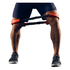
If you need help with this, our Hip & Core System can help guide your rehab.  It includes both our Hip & Core Band and a 30 Day Knee Fix will help to lay out a plan. Click here for more info